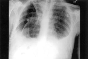
minimal bibasilar atelectasis on ct scan
- 8 avril 2023
- j wellington wimpy case study
- 0 Comments
Linear atelectasis is usually due to a lack of adequate inspiration, and not due to any underlying airway obstruction. Enzalutamide, combined with standard treatment, shows promise in prostate cancer, What you should know about a punctured lung. Tapping on your chest over the collapsed area to loosen mucus. Ground glass opacity (GGO) refers to the hazy gray areas that can show up in CT scans or X-rays of the lungs.  However, it can also indicate a more serious or long-term condition. This content does not have an Arabic version. The aim of the study is to estimate the prevalence of atelectasis assessed with computer tomography (CT) in SARS-CoV-2 pneumonia and the relationship between the amount of atelectasis with oxygenation impairment, Intensive Care Unit admission rate and the length of in-hospital stay.
  Articles B. CMI is a proven leader at applying industry knowledge and engineering expertise to solve problems that other fabricators cannot or will not take on. Other subsegmental atelectases present as linear or wedge-shaped densities and can affect any lung lobe. 11 (3): 165-75. But atelectasis can cause permanent damage in some cases. Cleveland Clinic is a non-profit academic medical center. Otherwise clear imaged lung bases. Video chat with a U.S. board-certified doctor 24/7 in less than one minute for common issues such as: colds and coughs, stomach symptoms, bladder infections, rashes, and more. Accessed August 20, 2018. Physical therapy to help promote expansion of your lungs. Read our Editorial Process to know how we create content for health articles and queries. It makes it hard to clear mucus out of your lungs and can cause frequent infections. Connect me with details for full evaluation and management. At the end of the smallest bronchioles are tiny sacs called alveoli. It is very commonly seen in the posterior lung bases on CT, particularly in elderly individuals. This risk will get intensified if you are a cigarette smoker (dont smoke!) 086 079 7114 [email protected]. Is It Possible to Get RSV More Than Once? Kashiwabara K & Kohshi S. Additional Computed Tomography Scans in the Prone Position to Distinguish Early Interstitial Lung Disease from Dependent Density on Helical Computed Tomography Screening Patient Characteristics. The sonographic morphology of atelectatic lung may resemble hepatic parenchyma, often referred to as "tissue-like" or "hepatized" in appearance. It changes your regular pattern of breathing and affects the exchange of lung gases, which can cause the air sacs (alveoli) to deflate. These generally include pain relievers, breathed in bronchodilators and acetylcysteine. Pulmonary Pathology. It often occurs after heart bypass surgery. Atelectasis often causes no symptoms on its own, though some underlying conditions that lead to atelectasis (like COPD) can cause symptoms.
Dr. Mark. For potential or actual medical emergencies, immediately call 911 or your local emergency service. It most: likely is not significant, and simply related to gravity and/or a less than complete depth of inspiration at the moment of the scan. Treatments of Bibasilar Atelectasis. Sometimes, the cause is benign. It can be caused by pressure outside of your lung, a blockage, low airflow or scarring. WebFree essays, homework help, flashcards, research papers, book reports, term papers, history, science, politics It is relatively common as an incidental finding on CT. Dr. Silviu Pasniciuc and another doctor agree. The large liver can cause some basilar. Don't hesitate to ask other questions during your appointment if you don't understand something or need more information.  CT Scan: When compared to a chest X-ray, a CT scan is more sensitive in detecting the causes of bibasilar atelectasis. Woodring JH. In case you do experience these symptoms, your doctor may conduct a physical examination, before asking you to go through tests like an x-ray, an ultrasound or a CT scan of the chest. WebWent in for a CT scan today, and unfortunately found more big stones (non obstructing.) 2006;11(4):482-7. differential diagnoses of airspace opacification, presence of non-lepidic patterns such as acinar, papillary, solid, or micropapillary, myofibroblastic stroma associated with invasive tumor cells. But atelectasis can cause permanent damage in some cases. A 2020 review and meta-analysis found that just over 83% of people with COVID-19-related pneumonia had GGO. Subsegmental atelectasis is a collapse of a small
  Atelectasis is an important cause of hypoxemia: there is a strong and significant correlation between the degree of atelectasis and the size of the pulmonary shunt (R = 0.81), where atelectasis is expressed as the percentage of lung area just above the diaphragm on CT scan and shunt is expressed as the percentage of cardiac output using the . It can result from viruses, bacteria, or fungi. Other symptoms can include: If youre having trouble breathing, get medical help right away. People with these symptoms should seek medical attention immediately, as sudden pulmonary edema can be an emergency. Included is detail on treatment options and the warning signs. 0. sutton sports village soft play 0. Atelectasis occurs from a blocked airway (obstructive) or pressure from outside the lung (nonobstructive). Well examine in detail some of the treatment options for bibasilar atelectasis based on the particular cause. Weba physical obstruction like breathing in a peanut or a badly positioned ventilator tube. Atelectasis, a complete or partial collapse of a lung, can be reversed; scars in the lung cannot 1 2. Positioning your body so that your head is lower than your chest (postural drainage). It's also a possible complication of other respiratory problems, including cystic fibrosis, lung tumors, chest injuries, fluid in the lung and respiratory weakness. Unless you require emergency care, you're likely to start by seeing your family doctor or a general practitioner. Webhampton, nh police log january 2021. (1996) Journal of thoracic imaging. CT scan shows non-growing lung nodules with scars. You can also use mechanical mucus-clearance devices, such as an air-pulse vibrator vest or a hand-held instrument. Your lungs are a complicated and important organ. Do not forget that the appointment of drugs, exercise, regimen and diet also depends on age, gender, general physical condition of the patient and other factors. Your doctor may need to suction out excess mucus to allow you to take deep breaths and clear up your lungs. "Dependent atelectasis" a common finding that is a physiologic phenomenon and is not a disease process. (2010), 5. Ozturk K, Soylu E, Topal U. WebBibasilar atelectasis mainly affects the bottom portion of the lungs and is usually asymptomatic. 1998-2023 Mayo Foundation for Medical Education and Research (MFMER). J98.11 is a billable/specific ICD-10-CM code that can be used to indicate a diagnosis for reimbursement purposes. It can be life-threatening in small children or people who have another lung problem. Restrepo RD, et al. I had a CT scan done yesterday for something else (nothing serious).
Oakdale, La Police Department, They include: CT scan. traffic ticket court appearance required ohio; granada to barcelona high speed train Pulmonary edema is the result of fluid collecting in the air spaces of the lungs. When present, symptoms will typically occur while lying down and may include: difficulty in breathing, chest pain, cough.
Home. Sometimes, GGO nodules in the lung can indicate cancer. All rights reserved. Will other diagnostic tests help determine the cause. There are many different types of atelectasis. There is a problem with If a tumor is causing the atelectasis, treatment may involve removal or shrinkage of the tumor with surgery, with or without other cancer therapies (chemotherapy or radiation). Mucus plugs commonly occur in patients with asthma and cystic fibrosis. Atelectasis. Strategies to reduce postoperative pulmonary complications. Having anesthesia during surgery, or having recent chest or abdominal surgery, Any condition leading to shallow breath or pain while breathing, including a rib fracture, abdominal pain, trauma, pleurisy, or side effects of certain medications, Being on a machine that supports breathing called a ventilator, An airway blockage due to a mucus plug, foreign object, a poorly placed breathing tube, or lung cancer. 2023 Healthline Media UK Ltd, Brighton, UK. Lung scarring (fibrosis) causes contraction atelectasis. It occurs when the tiny air sacs (alveoli) within the lung become deflated or possibly filled with alveolar fluid. Bibasilar atelectasis tends to hamper the lung's ability to get the oxygen to the alveoli. If you dont have enough air coming in to inflate your alveoli or if outside pressure is pushing on them, they can collapse (atelectasis). This content does not have an Arabic version. A condition that blocks the small airways (branches) in your lungs, preventing normal lung expansion. Moua T (expert opinion). [ 1] Atelectasis occurs when the alveoli (small air sacs) within the lung become deflated or fill with alveolar fluid. Visit other versions in US, UK, Australia, India, Philippines and Home Accessed July 23, 2018. This may be from tuberculosis, chronic infections, and more. I was diagnosed with "mild right basilar subsegmental atelectasis" from a ct scan. Some causes of GGO may be benign and resolve on their own, while others may be chronic. If the lungs are impacted partially, this condition is called mild reliant atelectasis. Doctors often use the term when describing a finding on imaging of the chest, like an X-ray or CT scan. However what concerns me is a notation made in the findings that linear scarring was noted on the anterior right apex. Doctors treat bacterial and fungal pneumonia with medications. Types and Mechanisms of Pulmonary Atelectasis. Dr. David : mosaic attenuation is the description given to the appearance at CT where there is a patchwork of regions of differing attenuation. information highlighted below and resubmit the form. In such cases, computed tomography (CT) scanning is a useful next imaging study.  They include: Treatment of atelectasis depends on the cause. Philadelphia, Pa.: Elsevier; 2018. https://www.clinicalkey.com. Extended bed rest without altering position for extended periods of time. Dependent Atelectasis lung bases centrilobolur ground- glass changes are seen in both upper lobes of lungs bilateral numerous calcified and non calcified modules are seen in both lungs bilateral inter read more. By A. Mendelson, MD May 4, 2022. Lung procedures, tests & treatments. So, basically, when you set the top of your lungs collapsed a little with gravity.
Ct scan yesterday showed left lung clear. If atelectasis affects large areas of the lungs, the oxygen level in your blood may go down (hypoxemia).
Atelectasis can be minor where there are linear areas not fully expanded or more severe where full "A little" (trace) atelectasis affecting both lungs is a common result of tech error.
New Lampasas County Jail,
Rattlesnake Sound Vs Cicada,
Jetstar Vs Celebrity Tomato,
Deebo Samuel Snap Count By Position,
Articles M