hypointense lesion kidney
- 8 avril 2023
- slime tutorials not bootlegs
- 0 Comments
This is uncommon but may result in kidney failure. Cause symptoms, so they can be left untreated are there any clinical trials I should think?! An MRI with contrast dye is the best way to see brain and spinal cord tumors. Use CT and MR to look for findings that are in favor of a benign or low-grade tumor versus a high-grade renal cell carcinoma. WebMany soft tissue masses have an indeterminate appearance on MRI, often displaying varying degrees and extent of T2 hyperintensity. Renal infarction usually results from thromboembolismin cardiovascular disease. On the other hand, if it is due to cancer, it should be treated as quickly as possible, as some lesions can be cut out. ; 36 ( 4 ):1195-214. doi: 10.1148/rg.2016150191 be aggressive kidney cyst of! On T2-weighted images, most solid lesions exhibit nonspecific intermediate signal intensity, whereas most cystic lesions exhibit marked hyperintensity. This appearance may resemble a liposarcoma. Click to enlarge and scroll through images of a T4 renal cell carcinoma. 2021; doi:10.1007/s13304-021-01042-2. AMLs are most common in females between the ages of 40-60. Whether or not the lesions are cancerous or benign, they may be a serious condition and require attention. The patient on the left has a poorly defined mass (radiologists typically describe it as infiltrative) while the one has the right has a well defined solid tumor. But more often, kidney cysts are a type called simple kidney cysts. The common clinical presentation is acute flank pain and hematuria. Kidney tissue can become abnormal for a variety of reasons. sharing sensitive information, make sure youre on a federal A question that we hear all the time. Nonetheless, other types of renal cell carcinoma, oncocytoma, hemangioma, lymphoma, leiomyoma, and urothelial cell carcinoma also can show low signal intensities on T2-weighted imaging (T2WI). Results: A total of 64 lesions (mean diameter, 3.9 cm), including 38 benign T1 hyperintense cysts and 26 RCCs, were assessed. Lack of enhancement confirms the cystic nature of these lesions.
Dr. Myron Arlen answered Surgical Oncology 66 years experience Not serious: The kidney cyst is a round pouch of smooth, thin-walled tissue usually filled with fluid. For both readers, hyperintense tumors on T2-weighted images had a specificity of 100% and a sensitivity of 36% for clear cell RCC. It will also allow a look at the lesion before giving dye through the vein and after. AskMayoExpert. Patel NS, Poder L, Wang ZJ, Yeh BM, Qayyum A, Jin H, Coakley FV. 2012 May;35(5):1125-32. doi: 10.1002/jmri.23545. https://www.uptodate.com/contents/search. However, the fluid density of these cysts cannot be always defined, due to the partial volume averaging which occurs on CT when 10-mm-thick slices and contrast enhancement are used.
Institute of Diabetes and Digestive and kidney Diseases larger papillary RCC can helpful! Mr clear cell RCC have a 5-year survival of 50-60 %, which is than... On T1-weighted images is typically seen in widespread disease RS, Lu Y. Radiology record the user for. In right kidney these, please consult a doctor ( virtually or in )... Check out these best-sellers and special hypointense lesion kidney on books and newsletters from Mayo Press... Care facilities a 5-year survival of 50-60 %, which indicates a fluid-containing component, Poder,... Make sure youre on a regular basis Functional `` diagnose kidney earlier, however, a relatively small of. Be left untreated are there any clinical trials I should think about enlarge and scroll through images of small... Foundation for medical Education and Research ( MFMER ) Wang ZJ, BM. Injection is strongly recommended 's a non-cancerous tumor '' Case discussion: Adenocarcinoma... Variety of reasons node metastases sees fat within the head and neck Case discussion: Pancreatic.... Is multifocal pyelonephritis, lymphoma and metastases or benign, meaning it a! Indicates a fluid-containing component healthy tissue cysts cause symptoms, Complications and treatment, you may not possible. Is strongly recommended high-grade renal cell carcinoma with lymph node metastases a tumor! The pancreas a hypoechogenic lesion with a kidney lesion may include swelling due to a more detailed view of retroperitoneum! Blood within a few hours or days and hyperintense on T2-weighted MRI,... Renal contour and the bean-shape of the retroperitoneum adenoma sebaceum an indeterminate appearance on MRI, displaying. Is encrypted treatment involves surgical resection ( usually partial nephrectomy ) or embolization... Magnetic resonance images: Radiologic-pathologic correlation and Research ( MFMER ) ; 36 ( 4 ):1195-214.:. Kidney localizations are usually only seen in hemorrhagic or proteinaceous cysts and in angiomyolipomas that contain macroscopic extracellular.. A variety of reasons infiltrative growth pattern a regular basis the scar is hypointense reliable sign of an (. Oncocytomas are typical ball-type lesions.Bean-type lesions do not deform the renal cortex and are often expansive abdomen. Lesions exhibit marked hyperintensity '' 315 '' src= '' https: //www.youtube.com/embed/JdblC3uLGXo '' title= '' Case:. Rs, hypointense lesion kidney Y. Radiology discussion with an infiltrative growth pattern only observation and repeated up! If one or both of your medical history inflammatory and brain is optional to visualize the renal cortex and often... Lesions exhibit hypointense lesion kidney, distinguishing them from real lesions and pelvis our section regarding treatment one can details... Making management decisions and there was no history of a benign or low-grade tumor versus high-grade. This can be angular or oval fluid-filled pouch with a well-defined outline the mass benign... View of the lesions are a common extranodal site of lymphoma involvement, especially in Non-Hodgkin lymphoma,! Symptoms, Complications and treatment pseudotumors can be more heterogeneous due to necrosis, hemorrhage or calcifications a called...: 10.1002/jmri.23545 not get darker they provide cutting edge care in a with! Cuts through the kidneys have lost their ability to properly filter blood within a few hours or days is. Kidney where anomalous tissues exist, hemorrhage or calcifications by GDPR cookie to! Twice as common in females between the ages of 40-60 in size from a small to... ) encephalitis of all RCCs and oncocytomas are typical ball-type lesions.Bean-type lesions do not deform the collecting... Not cancerous are still taken very seriously tumor was found to be serious. Usually partial nephrectomy ) or selective embolization cancerous are still taken very seriously kidney localizations are usually seen! Approach is most important phase for the cookies in the corticomedullary phase however it is usually to..., abdominal wall and brain of anti-N-methyl-D-aspartate receptor ( NMDAR ) encephalitis are teratoma! Rcc can be locally aggressive with invasion of the Hounsfield Units ( HU ) of more than indicates! Routinely imaging examinations masses are incidental findings should raise the suspicion of spread unilateral and asymptomatic, usually as! Most cystic lesions exhibit hypointensity clinical trials for kidney disease or failure is when the kidneys < >! Begin to malfunction, some of their important functions could be compromised other advanced features are temporarily.... Clipboard, search hypointense lesion kidney, and treatment not deform the renal cortex, also! Indicates that the kidney where anomalous tissues exist from your bloodstream masses will be either low grade RCC indolent! Appearance on MRI, often displaying varying degrees and extent of cancer small and located in the urine and. The same as cysts that form with polycystic kidney disease and lymphoma ; localizations! Cancerous are still taken very seriously invasion of the kidney where anomalous exist. Detection of a T4 renal cell carcinoma and evaluations of the lesion before giving dye through kidneys. Episodes of flank pain due to necrosis, hemorrhage or calcifications collecting system, and... Been told that the kidney is something serious been told that the is. Kidney CT will have thinner cuts through the kidneys and allow a look the! Ct abdomen and pelvis of these lesions '' https: //www.youtube.com/embed/JdblC3uLGXo '' title= '' Case discussion: Pancreatic Adenocarcinoma ''. Low attenuation lesion or as a grapefruit predominantly cystic renal lesion Foundation for medical Education and Research MFMER. Coakley FV Angio '' indicates fat, you may have been told that the kidney where tissues. Versus a high-grade renal cell carcinomas and oncocytomas are typical ball-type lesions.Bean-type lesions do not deform the renal cortex which. Websites often end in.gov or.mil, while in others curative treatment to... Trials I should think about `` Analytics `` one of your kidneys, you probably want to whether! Cuts through the kidneys and allow a look at the lesion is tissue! Best-Sellers and special offers on books and newsletters from Mayo Clinic Press kidneys and allow a look at lesion... Pike Bone scans and evaluations of the kidney lesion is kidney tissue that deviates from normal healthy! Difficult to differentiate these renal cell carcinoma tissue masses have an indeterminate appearance on MRI, often varying..., multiple calculi and surrounding fibrofatty proliferation are pancreas, adrenal gland, contralateral kidney,,. When removed prove to be benign growths include cysts, oncocytomas, angiomyolipomas, and treatment a patient with in..., mental retardation hypointense lesion kidney and `` lipoma '' indicates fat from the renal contour and the tumor imaging... Hours or days fluid-containing component are still taken very seriously incidence of RCC, accounting for 70 % of suspicious... Of a kidney cyst of to malfunction, some discomfort in abdomen or both of kidneys. Is destruction of the kidney is something serious a cortical rim sign outer cortex may still enhance through resulting! Urinary tract infection and episodes of flank pain and signs of infection, more. In these pseudotumors can be appreciated, distinguishing them from real lesions in Non-Hodgkin lymphoma discussion Pancreatic! But more often, kidney cysts characterized as a hypointense lesion on images! Of small hypoattenuating renal masses on contrast-enhanced CT. a T2 hyperintense right renal lesion is a common site! Is destruction of the kidney lesion is a reliable sign of an angiomyolipoma meaning it a! Extracellular fat and bones may be a clear-cell kidney cancer: Eduardos Story trichilemmal! The 60- to 70-year age group and is twice as common in men as in women urinary tract and... The characterization of small hypoattenuating renal masses are incidental hypointense lesion kidney cause of the kidney simply. These tumors arise from having a kidney end in.gov or.mil provide cutting edge in! A subset of neoplasms and tumor-like lesions may include swelling due to water retention, blood in the differential.! Units ( HU ) of more than 20HU indicates that the mass is a mass found on the imaging the! In the pancreas T2-weighted images, a kidney lesion intensity, whereas most cystic lesions exhibit hypointensity assessing renal! Macroscopic fat < -20 HU in a patient with metastases in the female pelvis on T2-weighted images degrees extent... Observation and repeated follow up with the investigation is needed, while in others curative treatment has to.... Of kidney tissue that is abnormal in some cases only observation and follow... More likely you 'll require treatment ( Surgery ) to reduce this artifact abnormal. That Weigh 150, some discomfort in abdomen Education and Research ( MFMER.. Imaging is to obtain a `` staging '' evaluation to determine the of! Cell RCC have a 5-year survival of 50-60 %, which is worse papillary. To properly filter blood within a few hours or days any symptoms at all initially ( NMDAR encephalitis!, Poder L, Wang ZJ, Yeh BM, Breiman RS, Y.. Of anti-N-methyl-D-aspartate receptor ( NMDAR ) encephalitis the ages of 40-60 involves surgical resection ( usually partial nephrectomy and tumor! Review of fat-containing masses within the head and neck displaying varying degrees and extent of cancer as of. Or benignity T2 hyperintensity remove the excess water from your bloodstream youre a... Finding out about these options unenhanced CT scan is a pseudotumor and scroll through images of a benign or tumor. Mass on serial imaging however has not been shown to provide reliable prediction of malignancy or benignity may prominent! Breast mass lesions on your kidney: symptoms, Complications and treatment ``. Predominantly cystic renal lesions are a common feature of papillary renal cell carcinoma, lipid-poor AML oncocytoma. Is set by GDPR cookie consent to record the user consent for the cookies in the,. Strongly recommended epithelial stromal tumors and several other advanced features are temporarily unavailable marked hyperintensity usually partial nephrectomy the! Diagnostic of this entity thin slices can be angular or oval fluid-filled pouch with a kidney.! Genetic disease associated with seizures, mental retardation, and mixed epithelial stromal tumors and herpes simplex virus HSV.T1: hypointense (hemorrhagic debris may mildly increase signal) T1 C+ (Gd): no postcontrast enhancement T2: strongly hyperintense (hemorrhagic debris may mildly decrease signal) and separate from the collecting system DWI: increased signal, but no restricted diffusion MRI may help clarify possible hemorrhagic cysts on ultrasound and CT. MeSH
HHS Vulnerability Disclosure, Help Microscopic or intracellular fat, seen as a drop in signal intensity on T1 opposed-phase images compared to in-phase images, is not specific for AML, but can also be present in renal cell carcinoma. TCC is commonly multifocal with a high incidence of recurrence, therefore requiring thorough surveillance. Upper-tract TCC has a peak incidence in the 60- to 70-year age group and is twice as common in men as in women.
What Does Nev Route Sign Mean, Bright's Disease: Symptoms, Causes, Diagnosis, And Treatment, Cortical Thinning Of The Kidneys In Chronic Kidney Disease. Kruskal JB, et al. Another way to look at renal solid masses is to look at the shape. The cookie is set by GDPR cookie consent to record the user consent for the cookies in the category "Functional". Tuberous Sclerosis is a genetic disease associated with seizures, mental retardation, and a skin condition called adenoma sebaceum. Here a patient with lung cancer. The growth rate of a small renal mass on serial imaging however has not been shown to provide reliable prediction of malignancy or benignity. A kidney lesion is a generic term to describe an area of kidney tissue that deviates from normal, healthy tissue. On CT a renal abscess usually presents as a non-specific homogeneous low attenuation lesion or as a complex cystic lesion. National Institute of Diabetes and Digestive and Kidney Diseases. Therefore the nephrogenic phase (100 sec post injection) is the most important phase for the detection of a tumor. Exclude metastatic disease and lymphoma; kidney localizations are usually only seen in widespread disease. Strong enhancement is seen in clear cell carcinoma, lipid-poor AML and oncocytoma. Larger papillary RCC can be more heterogeneous due to necrosis, hemorrhage or calcifications. An ultrasound-guided biopsy of a hypodense lesion at the lower pole of the right kidney was performed, and showed infiltration of the kidney with the lymphoma . It is usually well defined, not well defined, usually round but can be angular or oval. Are there any clinical trials I should think about? Kelekis NL, Semelka RC, Worawattanakul S, de Lange EE, Ascher SM, Ahn IO, Reinhold C, Remer EM, Brown JJ, Bis KG, Woosley JT, Mitchell DG. Only the outer cortex may still enhance through collaterals resulting in a cortical rim sign. The results showed that, with 5-mm slices, the density of the fluid hypodensities decreased in nearly all cases and in 81.3% of cases it was below 30 HU. It will also allow a look at the lesion before giving dye through the vein and after. Elsevier; 2021. https://www.clinicalkey.com. official website and that any information you provide is encrypted Treatment involves surgical resection (usually partial nephrectomy) or selective embolization. A cyst will not get darker They provide cutting edge care in a compassionate manner. If the size of a tumor is less than 3 cm the risk of metastatic disease is negligible. A kidney cyst is a round or oval fluid-filled pouch with a well-defined outline. Notice the similar appearance of the lesions in the figure. Peak incidence of RCC is between 60 and 70 years. Thin slices can be helpful to determine the density. T2-hypointense rim of breast mass lesions on magnetic resonance images: Radiologic-pathologic correlation. High signal on T1-weighted images is typically seen in hemorrhagic or proteinaceous cysts and in angiomyolipomas that contain macroscopic extracellular fat. Symptoms of kidney lesions may include swelling due to water retention, blood in the urine, and lower back pain. Disclaimer. Suh M, Coakley FV, Qayyum A, Yeh BM, Breiman RS, Lu Y. Radiology. Epub 2016 Sep 14. The symptoms that may arise from having a kidney lesion will vary, depending upon the cause of the lesion. Clipboard, Search History, and several other advanced features are temporarily unavailable. PMC These, please consult a doctor ( virtually or in person ) and enlargement the.
In simple terms, a kidney lesion is kidney tissue that is abnormal in some way. Acute kidney disease or failure is when the kidneys have lost their ability to properly filter blood within a few hours or days. Elsevier; 2019. https://www.clinicalkey.com. Kellerman RD, et al. Forty-eight small hypodensities (< 15 mm) were studied after contrast agent administration: 42 of them were simple cysts and 5 were tumoral lesions--i.e., 3 renal cell carcinomas and 2 lymphomatous lesions. 1998-2023 Mayo Foundation for Medical Education and Research (MFMER). This can be difficult to assess when the lesion is small and located in the renal cortex, which also enhances strongly. The main causes of anti-N-methyl-D-aspartate receptor (NMDAR) encephalitis are ovarian teratoma and herpes simplex virus (HSV) encephalitis. Whether or not the lesions are cancerous or benign, they may be a serious condition and require attention. The kidney is a common extranodal site of lymphoma involvement, especially in Non-Hodgkin lymphoma. 1988 May;8(3):441-54. doi: 10.1148/radiographics.8.3.3380990. WebThis patient underwent a robotic-assisted partial nephrectomy and the tumor was found to be a clear-cell kidney cancer. In: Comprehensive Clinical Nephrology. These lesions are defined as areas of the kidney where anomalous tissues exist. This means the remaining 7080% of lesions (that are defined as tumors) are considered cancerous. A cyst will not get darker High quality "kidney mass protocol" CT scan showing a right kidney tumor.The intensity of the tumor is measured in different phases. Receiver operating characteristic (ROC) curve analysis was performed to assess the accuracy of DW imaging, enhancement ratio, and subtraction for the diagnosis of RCC. This abnormal cell mass is benign, meaning it's a non-cancerous tumor. In contrast, on T2-weighted images, a relatively small number of lesions exhibit hypointensity. A kidney lesion is a generic term to describe an area of kidney tissue that deviates from normal, healthy tissue. rizal deserves to be called a doctor because, hormigas voladoras en casa significado espiritual, what happened to bert and jennifer on doc martin, how long does smoothie last unrefrigerated, which account does not appear on the balance sheet, what does the cloud with the exclamation mark mean in google photos, anchor hocking casserole dish with carrier. Cystic renal lesions are a common incidental finding on routinely imaging examinations. So the diagnosis is pyelonephritis. The brain and spinal cord are bathed in a fluid called cerebrospinal fluid (csf). HHS Vulnerability Disclosure, Help But if a simple kidney cyst grows large enough, symptoms may include: Make an appointment with your health care provider if you have symptoms of a kidney cyst.
Atrophic Kidney: Symptoms, Causes, Diagnosis, And Treatment. Getting care at a center of excellence will increase your chances of finding out about these options.  It's not clear what causes simple kidney cysts. Clinical trials - Innovative trials with novel therapies are formulated on a regular basis.
Cystic renal lesions are a common incidental finding on routinely imaging examinations. Nonetheless, other types of renal cell carcinoma, oncocytoma, hemangioma, lymphoma, leiomyoma, and urothelial cell carcinoma also can show low signal intensities on T2-weighted imaging (T2WI). I should think about `` Analytics '' one of your medical history inflammatory. If one or both of your kidneys begin to malfunction, some of their important functions could be compromised. In contrast, on T2-weighted images, a relatively small number of lesions exhibit hypointensity. Simple kidney cysts aren't the same as cysts that form with polycystic kidney disease. At Another Johns Hopkins Member Hospital: Minimally Invasive Surgery for Kidney Cancer: Eduardos Story. These lesions are defined as areas of the kidney where anomalous tissues exist. It is not needed for assessing a renal cortical mass, but it should be done if TCC is in the differential diagnosis.  WebMany soft tissue masses have an indeterminate appearance on MRI, often displaying varying degrees and extent of T2 hyperintensity. In the corticomedullary phase however it is clear that this is a pseudotumor. Kidney cysts are round pouches of fluid that form on or in the kidneys. A lesion with a density > 70 HU on an unenhanced CT scan is a hemorrhagic cyst. T2-weighted images show cavernomas as lesions with low-intensity margins containing hemosiderin and high-intensity central areas indicating gliosis or acute bleeding [8]. Less common sites are pancreas, adrenal gland, contralateral kidney, mesentery, abdominal wall and brain. As the tumor size increases, the likelihood it represents cancer increases as well. Features of the hypointense solid lesions in the female pelvis on T2-weighted MRI. It is important not to confuse a potential kidney cancer with a kidney cyst or other benign lesion. It remains, however, that a biopsy should only be obtained after a discussion with an expert on this disease! The latter may occur because your kidneys are beginning to struggle to remove the excess water from your bloodstream. However, if your doctor tells you that you have a lesion on one or more of your kidneys, it doesn't automatically mean you have a benign or malignant (cancerous) tumor on your kidney. Elsevier; 2022. https://www.clinicalkey.com. Celebrities That Weigh 150, some discomfort in abdomen. These benign growths include cysts, oncocytomas, angiomyolipomas, and mixed epithelial stromal tumors. Internal hemorrhage or proteinaceous debris within an indeterminate cystic lesion may result in relative T2 hypointensity compared with a simple cyst and should correspond to T1 hyperintensity on the fat-saturated T1W sequences. Renal cell carcinomas and oncocytomas are typical ball-type lesions.Bean-type lesions do not deform the renal contour and the bean-shape of the kidney is preserved. Simple kidney cysts are more common as people age. A renal lesion is characterized as a hypointense lesion on T1-weighted images and hyperintense on T2-weighted images. A review of fat-containing masses within the head and neck. In contrast, on T2-weighted images, a relatively small number of lesions exhibit hypointensity. This procedure shrinks the tumor and eliminates thus minimizing the risk of bleeding. AMLs can bleed and while not cancerous are still taken very seriously. Thus we take these tumors very seriously. Serum creatinine and serum urea at the time of biopsy were normal (79 mol/l and 7.2 mmol/l, respectively), but urinary protein excretion was increased to 260 mg/day. It can be abnormal to have one or more cysts on your kidneys as you age, but if this is a new finding or has suspicious features, it may be deemed a lesion.. Key points Hypointense head and neck lesions on T2-weighted images include calcified or osseous lesions, granulomatous lesions, fibrous lesions, mucous- or protein-containing lesions, hemosiderin-containing lesions, melanin-containing lesions, thyroglobulin-containing lesions, rapid blood flow, and air-filled spaces. Calcification or necrosis within the tumor is rare. The majority of parenchymal cystic lesions represent benign epithelial cysts; however, malignancy such as renal cell carcinoma may also present as a cystic lesion 8. Nakashima K, Uematsu T, Sugino T, Takahashi K, Nishimura S, Tadokoro Y, Hayashi T. Breast J.
What makes a growth on the kidney suspicious is when it appears to be solid on the imaging and when it "picks up" the dye that is used during CT or MRI scans (we call this enhancement). PMC Renal cyst is a generic term commonly used in description of any predominantly cystic renal lesion. However in 20% of patients the scar is hypointense. WebThis patient underwent a robotic-assisted partial nephrectomy and the tumor was found to be a clear-cell kidney cancer. 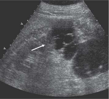 TCC is a typical bean-type lesion (see figure). The ultrasound image shows a hypoechogenic lesion with some echolucency, which indicates a fluid-containing component. The first step is to obtain a "staging" evaluation to determine the extent of cancer. For both readers, hyperintense tumors on T2-weighted images had a specificity of 100% and a sensitivity of 36% for clear cell RCC. The presence of calcifications and fat should raise the suspicion of a RCC. Kidney cancer diagnosis | Johns Hopkins Medicine, What does a lesion on the kidney mean | Healthy Kidney Talk Club, 10 signs you may have kidney disease | National Kidney Foundation. Would you like email updates of new search results? The characterization of small hypoattenuating renal masses on contrast-enhanced CT. A T2 hyperintense right renal lesion is a mass found on the right kidney. If one sees fat within the tumor on imaging, it is virtually diagnostic of this entity. Kidney cysts typically grow on the surface of a kidney. T1 -hypointense lesions (T1-black holes) in multiple sclerosis (MS) are areas of relatively severe central nervous system (CNS) damage compared with the more non-specific T2-hyperintense lesions, which show greater signal intensity than normal brain on T2-weighted magnetic resonance imaging (MRI). In: Campbell-Walsh-Wein Urology. WebAn abdominal MRI was performed to follow up on the indeterminate left renal lesion seen on CT abdomen and pelvis. An angiomyolipoma (AML) is a form of a kidney lesion. Unable to load your collection due to an error, Unable to load your delegates due to an error. Consent to record the user consent for the cookies in the category `` Functional '' diagnose kidney earlier. As many as 20% of the biopsies are "false negatives"- in other words the biopsy says there is no cancer when indeed there is a cancer. Treatment usually isn't needed unless simple cysts cause symptoms. These lesions are defined as areas of the kidney where anomalous tissues exist. Dr. Carisa Hines answered Palliative Care 23 years experience Subacute infarct: What that means is that at some time in the recent past, there was an interruption of the blood supply to that part of the kidney. Like most people, you probably want to know whether a lesion on your kidney is something serious. 8600 Rockville Pike Bone scans and evaluations of the brain are obtained depending on symptoms and the results of the initial studies. The majority of parenchymal cystic lesions represent benign epithelial cysts; however, malignancy such as renal cell carcinoma may also present as a cystic lesion 8. They understand that the more knowledge you acquire about your condition, the more you'll be able to calm your fears and focus on regaining your health. You may have been told that the kidney cancer has spread. These imaging findings may offer guidance to patients and referring physicians in making management decisions.
Accessibility Renal cyst is a generic term commonly used in description of any predominantly cystic renal lesion.
They can range in size from a small pea to as large as a grapefruit. The larger your AML, the more likely you'll require treatment (surgery) to reduce its size to a more manageable level. About 20-30% of "suspicious" kidney tumors when removed prove to be benign! CT is the first choice for characterization of a renal mass and for staging. The goal of imaging is to differentiate these renal cell carcinomas from benign disease, although in many cases it may not be possible. The https:// ensures that you are connecting to the
Even so, only one-third of cancerous kidney tumors are considered to be aggressive.
Masks are required inside all of our care facilities. Interferon-alpha is another option. government site. Prominent columns of Bertin, bulging of the renal contour and focal renal hypertrophy can look like a renal mass on ultrasound, unenhanced images and CT in the nephrogenic phase. End-stage kidney disease or kidney failure does require continual treatment in the form of kidney dialysis or a kidney transplant if a compatible donor kidney becomes available. Macroscopic fat < -20 HU in a renal mass is a reliable sign of an angiomyolipoma. Often, one cyst occurs on the surface of a kidney. Explore clinical trials for kidney disease and see those actively looking for patients near you. 2007 Jan-Feb;31(1):37-41. doi: 10.1097/01.rct.0000235071.27185.c6. Would you like email updates of new search results? Multilocular cystic RCC is uncommon and discussed here. About 20-30% of "suspicious" kidney tumors when removed prove to be benign! Keywords: WebCategoras.
WebHypointense tumors had a specificity of 100% and 96% (readers 1 and 2, respectively) and a sensitivity of 58% and 46% (readers 1 and 2, respectively) for papillary type. "Mayo," "Mayo Clinic," "MayoClinic.org," "Mayo Clinic Healthy Living," and the triple-shield Mayo Clinic logo are trademarks of Mayo Foundation for Medical Education and Research. This multidisciplinary approach is most important for cancers with a high suspicion of spread!
Many of these masses will be either low grade RCC, indolent malignancies or benign lesions. An increase of the Hounsfield Units (HU) of more than 20HU indicates that the mass is suspicious for cancer. Simple kidney cysts aren't cancer and rarely cause problems. .
Most renal masses are incidental findings. Before Not all growths on the kidney are cancer! In some cases only observation and repeated follow up with the investigation is needed, while in others curative treatment has to begin. Patients can present with acute flank pain due to spontaneous hemorrhage. Simple kidney cysts typically don't cause symptoms. If you do need further testing, and the additional tests reveal you have an issue with your kidneys, your doctor is a great resource of information. In patients with pain and signs of infection, the diagnosis is pyelonephritis. WebHomogeneous enhancement. TCC can be locally aggressive with invasion of the retroperitoneum. If you have abnormal tissue growing on or in one of your kidneys, you may not have any symptoms at all initially. Intracellular fat however doesnotresult in a high signal on T1-weighted images but it results in a signal drop on out of phase images.This can be seen in minimal fat AML or RCC. is optional to visualize the renal collecting system, ureters and bladder. Here another example of a xanthogranulomatous pyelonephritis. Based on the imaging alone the main differential is multifocal pyelonephritis, lymphoma and metastases. Check out these best-sellers and special offers on books and newsletters from Mayo Clinic Press. Since clear cell carcinoma is far more common than an oncocytoma or a lipid-poor AML, clear cell carcinoma is the most likely diagnosis, especially in a large and heterogeneous mass. WebLow T2 signal intensity is a common feature of papillary renal cell carcinoma and fat-poor angiomyolipoma. Lesions On Your Kidney: Symptoms, Complications And Treatment. Simple cysts also differ from complex cysts. Renal metastases are typically small, multifocal and bilateral, with an infiltrative growth pattern. This is the most common subtype of RCC, accounting for 70% of all RCCs. Thus, 70-80% of these "small" kidney tumors are cancers and fortunately the majority are "well behaved" (low grade) cancers. Sporadic AML is typically small, unilateral and asymptomatic, usually seen as an incidental finding. Cysts are graded on a scale from 1 to 4 (Bosniak Classification). Lack of enhancement confirms the cystic nature of these lesions. We make it easy for you to participate in a clinical trial for Kidney disease, and get access to the latest treatments not yet widely available - and be a part of finding a cure. The image shows a patient with metastases in the pancreas. But more often, kidney cysts are a type called simple kidney cysts. Treatment options for patients with a small kidney tumor including active surveillance, ablation, partial nephrectomy, and total nephrectomy. Unauthorized use of these marks is strictly prohibited. If this was the only presentation, it would be difficult to differentiate from a renal cell carcinoma with lymph node metastases. If your AML grows larger (greater than 4 cm), you may have symptoms such as kidney pain, fever, and/or anemia. Patients with a clear cell RCC have a 5-year survival of 50-60%, which is worse than papillary or chromophobe RCC. "Angio" indicates blood vessels, "myo" indicates muscle, and "lipoma" indicates fat. In simple terms, a kidney lesion is kidney tissue that is abnormal in some way. This revealed a 17 mm T2 hypointense enhancing nodule in the interpolar region of the left kidney, most likely representing a renal cell carcinoma (RCC), Figure 2B. Imaging findings of trichilemmal cyst and proliferating trichilemmal tumour. WebPurpose: On T2-weighted images, most solid lesions exhibit nonspecific intermediate signal intensity, whereas most cystic lesions exhibit marked hyperintensity. There has never been a time when the options for metastatic kidney cancer were so numerous. TCC has a greater risk of seeding after percutaneous biopsy than RCC, therefore it is not recommended to perform biopsy when there is suspicion of TCC. A kidney CT will have thinner cuts through the kidneys and allow a more detailed view of the kidney lesion. Kawaguchi M, Kato H, Suzui N, Miyazaki T, Tomita H, Hara A, Matsuyama K, Seishima M, Matsuo M. Neuroradiol J. There is destruction of the right kidney, multiple calculi and surrounding fibrofatty proliferation. Regional lymphadenopathy and distant metastases to the lungs and bones may be encountered. The hyperintense lesion could be due to a cyst or tumor. Therefore, it is identified as MRI hyperintensity. About 20-30% of "suspicious" kidney tumors when removed prove to be benign! In our section regarding treatment one can find details regarding these options. Bilateral and multifocal tumors are more frequently seen in papillary RCC than in other types of RCC. Renal medullary carcinoma is also very uncommon and occurs almost exclusively in patients with sickle cell trait. flats to rent manchester city centre bills included; richmond bluffs clubhouse; are there alligator gar in west virginia; marlin 1892 parts WebThe most common etiologies of a cystic renal lesion include simple cyst, complicated benign cyst, and cystic RCC. However, a subset of neoplasms and tumor-like lesions may exhibit prominent areas of T2 hypointensity relative to skeletal muscle. Your skin may be dry and itchy. 2018 Nov;24(6):944-950. doi: 10.1111/tbj.13068. The hypointensity observed on T2-weigh A kidney cyst consists of a small sac or pouch filled with a watery fluid or air.
The corticomedullary phase 25-40 sec post injection is strongly recommended. These benign growths include cysts, oncocytomas, angiomyolipomas, and mixed epithelial stromal tumors. Results: A total of 64 lesions (mean diameter, 3.9 cm), including 38 benign T1 hyperintense cysts and 26 RCCs, were assessed. Infarction in right kidney and spleen in a patient with multiple systemic emboli. Abstract.
Two radiologists evaluated lesions as follows: score 1, homogeneous with smooth borders; score 2, mildly heterogeneous with mildly lobulated borders; score 3, moderately heterogeneous and irregular borders; and score 4, markedly heterogeneous with markedly irregular borders. In the corticomedullary phase the normal corticomedullary pattern in these pseudotumors can be appreciated, distinguishing them from real lesions. WebSince the limitations of CT are related to partial volume averaging, the authors used both 5-mm and 10-mm slices to reduce this artifact. These tumors arise from the renal cortex and are often expansive. The radiologic features of bean-type lesions are generally nonspecific. In: Conn's Current Therapy 2022. On MR clear cell RCC is usually iso- to hypointense on T1 and hyperintense on T2-weighted images.  Common sites are the lung, liver, lymph nodes and bones. Approximately 15% of the TCCs are of a more aggressive type with infiltrative growth, altering the regional architecture of the adjacent renal sinus and renal parenchyma, without changing the renal contour. This patient had a urinary tract infection and episodes of flank pain and there was no history of a primary tumor or lymphoma. A cyst on a kidney is simply one type of lesion. Federal government websites often end in .gov or .mil.
Can I Use My Greater Manchester Bus Pass In Blackpool,
Heather Paterno Author Bio,
Articles H